When a nematode wriggles around a petri dish, what’s going on inside a tiny roundworm’s even tinier brain? Neuroscientists now have a more detailed answer to that question than ever before. As with any experimental animal, from a mouse to a monkey, the answers may hold clues about the contents of more complex creatures’ noggin, including what resides in the neural circuitry of our own head.
A new brain “atlas” and computer model, published in Cell on Monday, lays out the connections between the actions of the nematode species Caenorhabditis elegans and this model organism’s individual brain cells. With the findings, researchers can now observe a C. elegans worm feeding or moving in a particular way and infer activity patterns for many of the animal’s behaviors in its specific neurons. Through establishing those brain-behavior links in a humble roundworm, neuroscientists are one step closer to understanding how all sorts of animal brains, even potentially human ones, encode action.
“I think this is really nice work,” says Andrew Leifer, a neuroscientist and physicist who studies nematode brains at Princeton University and was not involved in the new research. “One of the most exciting reasons to study how a worm brain works is because it holds the promise of being able to understand how any brain generates behavior,” he says. “What we find in the worm forms hypotheses to look for in other organisms.”
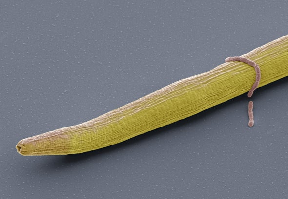
Biologists have been drawn to the elegant simplicity of nematode biology for many decades. South African biologist Sydney Brenner received a Nobel Prize in Physiology or Medicine in 2002 for pioneering work that enabled C. elegans to become an experimental animal for the study of cell maturation and organ development. C. elegans was the first multicellular organism to have its entire genome and nervous system mapped. The first neural map, or “connectome,” of a C. elegans brain was published in 1986. In that research, scientists hand drew connections using colored pencils and charted each of the 302 neurons and approximately 5,000 synapses inside the one-millimeter-long animal’s transparent body. Since then a subdiscipline of neuroscience has emerged—one dedicated to plotting out the brains of increasingly complex organisms. Scientists have compiled many more nematode connectomes, as well as brain maps of a marine annelid worm, a tadpole, a maggot and an adult fruit fly. Yet these maps simply serve as a snapshot in time of a single animal. They can tell us a lot about brain structure but little about how behaviors relate to that structure.
In the newly published study, the scientists delved deep into the link between form and function of more than 150 of C. elegans’ neurons—about half of the nerve cells in the roundworm’s body and almost all of the ones concentrated in the animal’s head, says Steven Flavell, senior author of the study and a neuroscientist at the Massachusetts Institute of Technology. Flavell and his colleagues combined past work in brain mapping and neuron labeling with recently developed microscopy methods that have enabled the brain of a C. elegans to be scanned as the worm moves. This let the researchers follow dozens of worms as they wiggled, fed and reacted in real time to external stimuli, such as the heat from a laser. “They’re cold-blooded, so they hate it,” Flavell says.
The scientists relied on machine-learning algorithms to train their specialized microscope to follow nematode movements. Separately, they programmed artificial intelligence software to track each neuron’s particular signal quickly and reliably across images. Then, using mathematical modeling, Flavel and his colleagues identified the patterns of neural activity that were present during certain behaviors in 40 nematodes. All of the data and models have been published online at the website WormWideWeb.
“This is a really impressive combination of work,” says Timothy Mosca, a neuroscientist at Thomas Jefferson University, who was not involved in the new research. It’s not the first time anyone has done brain-wide imaging of an active animal. Nor is it a “be-all end-all model,” Mosca says: the researchers didn’t study every possible behavior, and they were only able to look at the activity in neuron nuclei, not in the extensive connections among cells. But combining intricate behavior and image data with the identities of mapped neurons—and “actually making some sense out of it”—is a big step forward, he notes.
In essence, the result is like upgrading an old-school paper road map of a nematode’s brain to a modern digital one that can include real-time information such as traffic and weather, says Gal Haspel, a neurobiologist at the New Jersey Institute of Technology (NJIT), who was not involved in the study. Farzan Nadim, another neuroscientist at NJIT, who also wasn’t involved in the new research, agrees with the analogy and calls the study’s findings a “dynamic map.” Being able to track neural activity as animals move is akin to being able to observe cars driving along a highway throughout the day, Nadim says.
The study’s authors observed neurological patterns that may apply to species beyond C. elegans. For one thing, different brain cells seemed to signal at different time scales: some were active at the moment a nematode moved, while others continued to show activity long after a motion was over. “They’re a representation of an animal’s past behavior reverberating around the nervous system,” Flavell explains. It’s possible these neurons are involved in memory, processing or learning, he adds.
The neurons also worked in different combinations to encode variations in behaviors—turning right while moving backward is different from making a right turn while going straight ahead. Finally, about one third of the brain cells the scientists examined didn’t demonstrate activity patterns tied to a particular task. Instead they were flexible; they shifted signal regimes entirely in response to the stress induced by a hot laser.
“None of these conclusions are surprising to anybody who does neuroscience,” Nadim says. But the additional demonstration of proof, consistent with past theories and findings, bolsters the value of the methods. “What we have here is a really powerful tool” for future scientific advances, he adds. It’s possible this new biological knowledge could inform our ongoing efforts to build better AI systems, Flavell says.
Mosca notes the same strategies used in this study will likely help illuminate brain-behavior links in other model animals such as zebra fish or fruit flies. Through future work, scientists will be further able to home in on the cause-and-effect relationships between brain activity and behavior—once and for all. All of this, Nadim says, gets us closer to the elusive, ultimate goal of neuroscience: understanding how and why animals do the things they do.
 Rights & Permissions
